

Heritable Features of the Optic Disc:
A Novel Twin Method for Determining Genetic Significance
Purpose: Numerous genetic diseases and environmental stimuli affect optic nerve morphology. The purpose of this study was to identify the principal heritable components of visible optic nerve head structures in a population-based sample of twins.
Methods: Fifteen optic nerve specialists viewed stereoscopic optic nerve head photographs (Stereo Viewer-II; Pentax Corp., Tokyo, Japan) from 50 randomly selected monozygotic or dizygotic twin pairs. Before viewing, each expert was questioned about which optic nerve head traits they believed were inherited. After viewing a standardized teaching set, the experts indicated which twin pairs they thought were monozygotic. Participants were then questioned about how their decisions were reached. A rank-ordered Rasch analysis was used to determine the relative weighting and value applied to specific optic nerve head traits.
Results: The proportion of twin pairs for which zygosity was correctly identified ranged from 74% to 90% (median, 82%) across the panel. Experts who correctly identified the zygosity in more than 85% of cases placed most weighting on shape and size of the optic disc and cup, whereas experts with the lowest scores placed greater weighting on the optic nerve head vasculature in reaching their decisions.
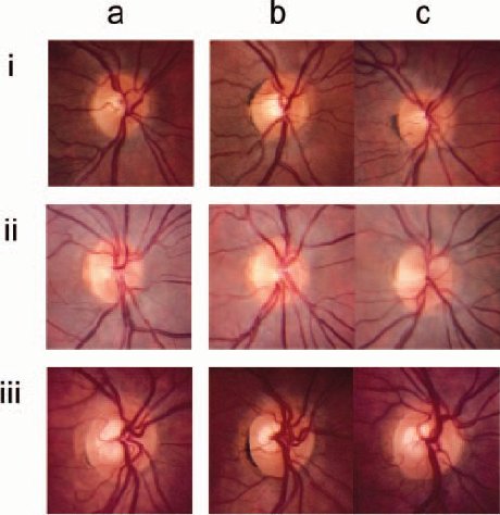
Figure 1. ONH photographs of MZ twin pairs for which each expert was asked, after viewing the right ONH from one of the MZ twin pairs (a), whether the same individual’s left ONH, flipped horizontally to appear as a right ONH, or the right ONH from the MZ twin, most resembled the first. In examples (i) and (iii), ONH photographs (a) and (c) were from the same individual, whereas in (ii), photographs (a) and (b) are from the same individual.
Conclusions: In determining the genetic components of the optic nerve head, the results of this study suggest that the shape and size of the optic disc and cup are more heritable and should receive a greater priority for quantification than should vascular features.
Invest Ophthalmol Vis Sci. 2007;48:2469–2475
Accepted for publication March 14, 2007
Alex W. Hewitt1,2, Johan P. Poulsen1, Wallace L. M. Alward3, Sonya L. Bennett4,
Wido M. Budde5, Richard L. Cooper6, Jamie E. Craig2, John H. Fingert3, Paul J. Foster4, David F. Garway-Heath4, Catherine M. Green7,7 Christopher J. Hammond8, Sohan S. Hayreh3, Jost B. Jonas5, Paul L. Kaufman9, Neil R. Miller10, William H. Morgan11, Nancy J. Newman12, Harry A. Quigley10, John R. Samples13, George L. Spaeth14,
Konrad Pesudovs2, and David A. Mackey1,15
1 Centre for Eye Research Australia, University of Melbourne, Royal Victorian Eye and Ear Hospital, Melbourne, Australia;
2 National Health and Medical Research Council (NHMRC) Centre for Clinical Eye Research, Department of Ophthalmology, Flinders University, Flinders Medical Centre, Adelaide, Australia;
3 Department of Ophthalmology, University of Iowa, Iowa City, Iowa;
4 Glaucoma Research Unit, Moorfields Eye Hospital, London, United Kingdom;
5 Department of Ophthalmology, Faculty of Clinical Medicine Mannheim, University of Heidelberg, Mannheim, Germany;
6 Tasmanian Eye Clinics, Launceston, Australia;
7 Glaucoma Research Unit, Royal Victorian Eye and Ear Hospital, Melbourne, Australia;
8 Twin Research and Genetic Epidemiology Unit, St. Thomas’ Hospital, London, UK;
9 Department of Ophthalmology and Visual Sciences University of Wisconsin (UW) School of Medicine and Public Health UW Clinical Science Center, Madison, Wisconsin;
10 Department of Ophthalmology, The Johns Hopkins Medical Institutions, Baltimore, Maryland;
11 Lions Eye Institute, University of Western Australia, Nedlands, Australia;
12 Department of Ophthalmology, Emory University School of Medicine, Emory Eye Center, Atlanta, Georgia;
13 Casey Eye Institute, Portland, Oregon;
14 William and Anna Goldberg Glaucoma Service and Research Laboratories, Wills Eye Hospital/Jefferson Medical College, Philadelphia, Pennsylvania;
15 Department of Ophthalmology, University of Tasmania, Royal Hobart Hospital, Hobart, Australia.
 Heritable Features of the Optic Disc
as PDF (463 Kb)
Heritable Features of the Optic Disc
as PDF (463 Kb)
![]()
Index of Papers
[
Welcome ][ Publications ]