

Terriens marginal degeneration: case reports and literature review
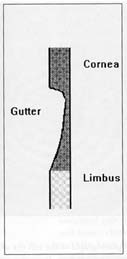
Terrien’s marginal degeneration (TMD) is a rare, bilateral, asymmetric disease of unknown aetiology. The peripheral cornea, predominantly superiorly, undergoes lipid deposition, vascularisation, opacification and stromal thinning leading to ‘gutter’ formation, ectasia and eventual corneal perforation. Two cases are presented which demonstrate the typical clinical features of the various stages of this disease. The disease process and its spectrum of presentation are reviewed. Differential diagnosis and management of TMD are discussed with particular reference to computerised corneal topographical analysis, which has a limited role for diagnosis but is valuable for monitoring disease progression.
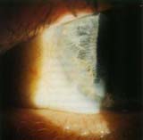
Figure 6. Eyesis polaroid photograph of the left eye of case two. Arrows denote
gross distortion of the peripheral cornea, from the guttering (closed) and over the pseudopterygium (open).
|
Clin Exp Optom 1994; 77: 97-104 Konrad Pesudovs BScOptom Accepted for publication: 3 February 1994 Key words: astigmatism, computerised corneal topographical analysis, ectasia, Terrien’s marginal degeneration. |
Figure 4. Pseudopterygium from the LE of case two. Note the broad, flat head and the oblique angle of insertion. |
|
Condition |
Differentiating features |
|
Pellucid marginal degeneration |
Inferior corneal thinning only; no pseudopterygia; inferior corneal ectasia only. |
|
Marginal furrow degeneration |
Asymptomatic, involves 360o of the cornea, no opacification, no vascularisation, no pseudopterygia, no ectasia, no change in astigmatism. |
|
Dellen |
localised corneal depression only, not extending circumferentially, adjacent to raised lesion, unlikely to be bilateral. |
|
Mooren's ulcer |
Inflammatory, epithelium is disrupted, with consequent fluorescein staining, tends to be localised, rapidly progressive. |
|
Rheumatoid disease |
Associated systemic disease, usually inflammatory, can have non-infiammatory peripheral gutter formation with vascuiarisation, opacity, lipid deposition, but progresses more rapidly and leads to keratolysis rather than keratectasia. |
|
Keratoconus |
No gutter, gross central/inferior ectasia, dramatic reduction of vision, often in young people, no peripheral opacification, no pseudopterygia. |
|
Fungal ulcer |
Inflammatory, localised, rapidly progressive, history of trauma. |
|
Acne rosecea |
Associated systemic disease, inflammatory more extensive pannus formation, loss of epithelial integrity. |
|
Arcus senilis |
Possible systemic hyperlipidaemia, no gutter formation, no vascularisation, no ectasia. |
Table 1. Differential diagnosis of Terrien’s marginal degeneration
 Article in full as PDF (900 Kb)
![]()
![]()
Index of Papers
[ Welcome ][ Publications ]