

Flicker perimetry and pigment epithelial detachment
We report a case of a 71-year-old man with a retinal pigment epithelial detachment (PED) and occult subretinal neovascularisation. Automatic perimetry was performed with a non-flickering stimulus and an 18 Hz flickering stimulus (flicker perimetry). The addition of flicker to the perimetric task enhanced the detection of the PED. It appears that testing the ability of the visual system to process temporally encoded information can enable detection of a deficit in visual function where there is a minimal defect for conventional increment thresholds.
|
Furthermore, given the known pathophysiology of PED, it appears that flicker perimetry can enable detection of a defect of photoreceptor function. Flicker perimetry may prove to be a useful tool for assessing retinal disease.
Clin Exp Optom 1994; 77: 58-63 Konrad,Pesudovs BSc Optom*, * Department of Ophthalmology, Flinders Medical Centre Accepted for publication: 8 December 1993
Key words: age-related maculopathy, flicker, perimetry, pigment epithelial detachment
|
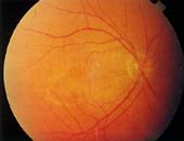
Figure 4. Fundus photograph of the right eye showing pigment epithelial detachment.
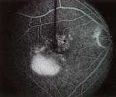
Figure 5. Fluorescein angiogram of the same eye (figure 4) showing a serous elevation with a small fleck of blood at the macula. This was interpreted by the retina specialist as an epithelial detachment with occult subretinal neovascular membrane formation within the foveal avascular zone. |
![]()
![]()
Index of Papers
[ Welcome ][ Publications ]